Why Are My Gums Pale or White? The 6 Patterns — and the Wipe-and-Spread Test That Shows Which Need a Doctor
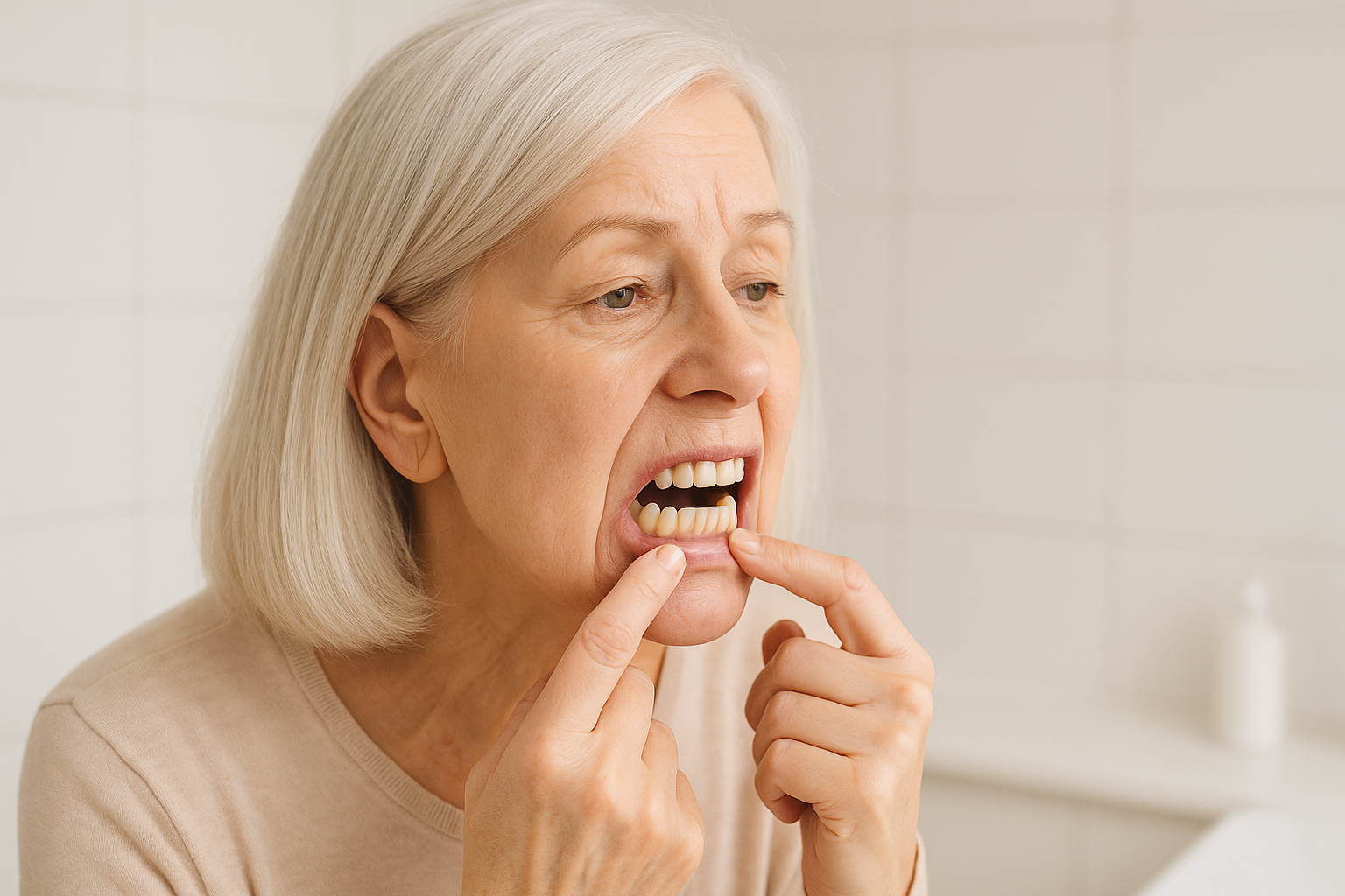
Last updated: 7 October 2026
Quick answer
Pale or white gums usually fall into one of two very different groups: the whole gum looks pale (a body-wide sign, classically low iron or anaemia) or distinct white patches or streaks sit on the gum (a mouth-surface problem such as thrush, oral lichen planus or leukoplakia). The Medical News Today clinical review names anaemia as the most common cause of pale gums, often from too little iron, folate or vitamin B-12. For patches, the single most useful clue is whether they wipe off: Mayo Clinic describes oral thrush as creamy white patches with “slight bleeding if the patches or spots are rubbed or scraped,” while leukoplakia is thick white patches that “cannot be scraped off.”
Three things should not be left to “see how it goes”: white patches that last more than two weeks, patches that cannot be wiped away, and gum pallor that comes with tiredness, breathlessness or a racing heartbeat. A dentist or doctor can settle the cause — and for anaemia only a blood test can. Nothing you apply to the gums changes a pale colour whose cause lies elsewhere; a water-free lipid gum-line concentrate such as Dental Pro 7 is one example of a cosmetic daily product that supports the look of healthier, pinker gums when the gum line itself needs care — it is not a response to pallor and does not replace a dental or medical check.
What “normal” gum colour actually is — and why it varies
Healthy gum tissue is pink because it is thin, richly supplied with blood vessels, and contains a variable amount of melanin — the same pigment that colours skin. That variability is the first thing to hold in mind: there is no single “correct” pink. Some people have naturally lighter, coral or even slightly pale gums; others have darker, melanin-rich gums (see why are my gums dark or purple?). Colour that has always been there and has not changed is rarely the issue.
What deserves attention is change and pattern. Pale gums that were pink last year, a lighter band that is new, or white areas with an edge you can point to are the situations worth sorting out. Because the causes range from harmless to “needs a blood test,” and the pages that currently rank for this question are mostly short lists of causes without a way to tell them apart, the decoder below is built around what you can actually observe.
The 6-pattern decoder
Match the description that fits your gums best. This layout is our own synthesis of the sources named in the table; it is a way to arrive at your dentist’s or doctor’s appointment with better information, not a diagnostic system.
| # | What you see | Most likely explanation | What the sources say | Who to see |
|---|---|---|---|---|
| 1 | Evenly light pink, symmetrical, always been this way, no symptoms | Natural variation | Gum colour depends on blood supply, tissue thickness and melanin — so it varies from person to person. (Our summary of normal anatomy; no single “normal” shade is defined in the sources below.) | Mention it at your next check-up; no urgency if long-standing and unchanged |
| 2 | The whole gum, and perhaps the lining of the mouth, looks washed-out or pale — plus tiredness, breathlessness, weakness, palpitations or pale skin | Anaemia (often low iron) | Medical News Today: “pale gums around the teeth may indicate anemia, which is often caused by a lack of iron, folate, or vitamin B-12.” StatPearls: iron-deficiency signs “can include pallor, dry mouth, atrophic glossitis, angular cheilitis, and hair loss,” but are “often subtle”; diagnosis rests on laboratory results, with serum ferritin below 45 ng/mL recommended for diagnosing iron-deficiency anaemia | GP / doctor — a blood-test question, not a mouth-product question |
| 3 | Creamy white, slightly raised patches “like cottage cheese” that wipe off leaving a red, raw or slightly bleeding surface; sore mouth, cracked corners, altered taste | Oral thrush (Candida overgrowth) | Mayo Clinic: patches on tongue and inner cheeks, sometimes the roof of the mouth, gums or tonsils, with “slight bleeding if the patches or spots are rubbed or scraped.” NHS: “When you wipe off the white patches, they leave red spots that can bleed.” Mayo notes thrush is “uncommon in healthy older children, teenagers and adults,” so a doctor may look for an underlying cause | Dentist or GP; NHS advice is to return if there is no improvement after 1 week of treatment |
| 4 | White lacy or web-like streaks (sometimes on the cheeks too), or red, raw, shiny gums that sting with spicy food and bleed on brushing | Oral lichen planus | Mayo Clinic: the reticular type “appears as white patches in the mouth and can look lacy”; the erosive type shows “red, swollen tissues or open sores”; gums are one site. StatPearls: about 10% of patients have lesions confined to the gingiva, as desquamative gingivitis; global prevalence about 2%; biopsy advisable to confirm the diagnosis | Dentist, then a specialist if needed; regular check-ups afterwards |
| 5 | Thick, firm white or grey patch that does not wipe off, may feel rough or ridged; often in a smoker or after years of alcohol use, or beside a broken tooth or poorly fitting denture | Leukoplakia | Mayo Clinic: patches “cannot be scraped off,” commonly on the gums, inner cheeks and floor of the mouth; tobacco is the leading cause, with alcohol, betel nut and physical irritation also named; most are benign but some show early cancer changes, and speckled white-and-red patches carry higher risk. See a provider if patches persist beyond two weeks | Dentist or doctor soon — do not wait it out |
| 6 | Pale, dry gums that coincide with menopause, or with known kidney problems | Hormonal or systemic change | Medical News Today attributes pale, dry gums in menopause to hormonal changes that reduce blood flow; Colgate lists kidney disease among causes of pale gums. Both are brief mentions rather than detailed mechanisms | Mention it to your GP or dentist as part of the wider picture |
Sources: Huizen J (med. rev. Luo EK), “Pale gums,” Medical News Today (upd. 26 Jun 2023); Jogu P & Kamran MT, “Iron-Deficiency Anemia,” StatPearls (upd. 15 Feb 2026); Mayo Clinic, “Oral thrush” (20 Dec 2024), “Oral lichen planus” (24 Dec 2024) and “Leukoplakia” (15 Feb 2024); StatPearls, “Oral Lichen Planus”; NHS, “Oral thrush (mouth thrush)”; Colgate Oral Care Center, “What causes pale gums.” The six-row layout is our synthesis.
Whole-gum pallor: why anaemia is the first thing to rule out
When the colour change is everywhere rather than in patches, the question stops being about your mouth and becomes a question about your blood. Anaemia means the blood carries less oxygen, and the Colgate Oral Care Center explains that people with anaemia “often” lack the iron needed to make haemoglobin, adding fatigue, an unusual heartbeat, weakness and pale skin to the picture. Medical News Today ranks it as the most common cause of pale gums and names iron, folate and vitamin B-12 as the usual shortfalls.
The honest limitation is that you cannot confirm this by looking. The StatPearls iron-deficiency chapter lists pallor among the possible findings but calls such signs “often subtle,” and says symptoms “may be absent or minimal in early or mild disease.” Its diagnostic anchor is a laboratory one: a serum ferritin below 45 ng/mL is recommended as the cut-off for diagnosing iron-deficiency anaemia. In practice that means: if your gums look washed-out and you are unusually tired, short of breath on stairs, or have noticed a pounding heartbeat or pale inner eyelids, ask for a blood test rather than buying a supplement first. Supplements started blind can mask the picture and, for iron, are not risk-free.
White patches: thrush, lichen planus or leukoplakia?
These three look similar in a mirror and are managed completely differently, which is why the next section matters. Here is the short version of each, using only what the opened sources state.
Thrush: white, creamy — and it wipes off
Mayo Clinic describes oral thrush as Candida albicans building up in the mouth: “creamy white patches” on the tongue and inner cheeks, sometimes the roof of the mouth, gums and tonsils, “slightly raised patches that look like cottage cheese,” with redness or soreness, a cottony feeling, loss of taste, and cracking at the corners of the mouth. A Healthwise article reproduced by HealthLink BC adds the useful visual: the patches look like cottage cheese or milk curds, and wiping them away reveals a red, raw surface that may bleed. Risk factors named by Mayo include dentures (especially upper), inhaled corticosteroids, antibiotics, poorly controlled diabetes and dry mouth. If antibiotics are the trigger, see why breath changes after antibiotics; if dry mouth is, see why is my mouth always dry?
Oral lichen planus: lacy streaks, or a raw red gum
Mayo Clinic says the cause is unknown, with activated T lymphocytes suggesting an immune element. The reticular form usually causes no discomfort; the erosive form brings burning or pain, sensitivity to hot, acidic or spicy foods, and “bleeding and irritation with toothbrushing.” Gums are a recognised site, and in StatPearls roughly one in ten patients have disease confined to the gums, presenting as desquamative gingivitis — a raw, red, peeling-looking gum rather than a white one, which is why people searching for “white gums” sometimes miss it. On risk, StatPearls cites a meta-analysis putting malignant transformation at 0.44% (lower than most earlier estimates), with higher risk in smokers, heavy drinkers, people with hepatitis C and the red subtypes; Mayo says regular check-ups are needed because the erosive type “may raise the risk of getting mouth cancer.” The balanced reading: usually manageable, worth confirming by biopsy, and worth monitoring.
Leukoplakia: the patch that will not come off
Mayo Clinic’s definition is blunt: thick white patches that “cannot be scraped off,” with a rough, ridged, wrinkled or smooth surface, mostly on the gums, inner cheeks and floor of the mouth. Tobacco is the leading cause. The reason it gets urgency is that it can be an early warning of cancer in a minority of cases, particularly speckled leukoplakia (white mixed with red). Mayo’s threshold is practical: see a provider if patches persist beyond two weeks, or if there are lumps, trouble swallowing or ear pain. A separate form, hairy leukoplakia, causes fuzzy white patches on the sides of the tongue and mostly occurs in people with weakened immunity.
The wipe-and-spread test
This is the information-gain piece: a way to sort your gums by what you can observe, so the appointment starts further along. It takes about five minutes on day one and two minutes on day 14. It is our synthesis of the sign descriptions above and is not a diagnosis.
- Look in daylight, not bathroom light. Photograph the front view, left and right views and a close-up of the paler or whiter area with a coin beside it for scale.
- Wipe, gently. After brushing, press a clean, damp gauze or tissue lightly over the white area for a second or two. Do not scrub. If the white comes away and leaves a red or slightly bleeding surface, that matches the descriptions of thrush (Mayo Clinic, NHS). If nothing comes off, that matches leukoplakia (“cannot be scraped off”) and is also true of lichen planus streaks — either way it is a dentist visit.
- Map it. Is the paleness everywhere (pointing toward blood and general health), a patch or two (a surface lesion), or a lacy network that may also sit on the inside of the cheeks (lichen planus)? Check the inner cheeks and tongue for the same colour.
- Tick the companions. New tiredness, breathlessness or palpitations; sore mouth or cracked corners; a new medicine, inhaler, antibiotic course or denture; smoking or regular heavy alcohol use; a burning feeling with spicy food.
- Re-photograph on day 14 in the same light with the same coin. Mayo’s two-week line for white patches is the benchmark: patches that persist beyond two weeks should be looked at. The Cleveland Clinic gives a similar two-week rule for gums that bleed.
- Book the appointment sooner if any patch cannot be wiped away, is rough, firm, red-and-white, growing, painful or bleeding without being touched, or if you have trouble swallowing. Take both sets of photos; they are far more useful than a description.
Why photographs: clinicians ask “has it changed?”, and memory is a poor instrument for slow change in a place you cannot see easily.
What to expect at the visit
For a possible thrush picture, expect questions about medicines, inhalers, dentures and diabetes, and a prescription antifungal if it is confirmed. For lacy or erosive patches, the StatPearls recommendation is a biopsy to confirm the diagnosis and rule out dysplasia, followed by long-term surveillance. For a patch that will not wipe off, a dentist will usually examine it, ask about tobacco and alcohol, and arrange a biopsy or referral if there is any doubt. For whole-gum pallor with tiredness, the route is your GP: a full blood count and iron studies including ferritin.
Where a gum-line lipid concentrate fits — and where it doesn’t
Being precise about the role matters here, because most of the patterns above sit outside what any cosmetic can address. Nothing applied to the gums changes anaemia, clears thrush, resolves lichen planus or alters a leukoplakia patch, and no product replaces having a persistent white area assessed. Where a daily gum-line product fits is the ordinary job of keeping the gum line looking its best between dental visits — the cleaning habits covered in how to brush with gum disease come first.
Dental Pro 7 is a 100% water-free botanical lipid concentrate built on a design principle we call Lipid-Lock: because it contains no water, it clings to the gum line and stays in contact for hours rather than being flushed away in seconds the way a water-based paste or rinse is. It blends eleven botanical lipids — immortelle helichrysum, pomegranate seed, black cumin seed, Indian myrrh, wild clove, white thyme and eucalyptus among the actives, a peppermint–spearmint–wild-mint freshness trinity, and a grapeseed, sunflower and vitamin E base — and supports the look of firmer, pinker, healthier-looking gums, with the look of redness reduced, and a mouth that looks and feels cleaner and fresher.
How to use it: four drops on a dry toothbrush in place of toothpaste, brush gently for about two minutes with the bristles angled into the gum line, then spit — do not rinse with water. Rinsing washes the lipid layer straight off and defeats the design. If you are in the middle of the wipe-and-spread test and have a sore, raw or bleeding area, pause and let a dentist look first rather than applying anything new to it.
Because the formula is anhydrous it is preservative-free by design — no water, no fillers, no SLS or foaming agents, no fluoride, no parabens; vegan and non-GMO. It naturally contains eugenol, limonene and linalool from its botanicals, and it is made in a facility that handles nut oils.
- Rating: Dental Pro 7 is rated 4.81 out of 5 from 190 verified buyers, with over 500,000 bottles sold.
- Guarantee: backed by a 90-day money-back guarantee.
- Formulated by S. C. Aris.
- What it is for: supporting the appearance of a cleaner, fresher mouth and healthier-looking gums. It is a cosmetic complement to brushing, interdental cleaning and professional dental care — not a substitute for any of them, and it does not change gum colour caused by anaemia, infection or a mouth-surface condition.
Explore Dental Pro 7. Companion reading: early gum disease: signs and self-check, why do my gums bleed?, why do my gums look like an orange peel? and understanding puffy gums.
Frequently asked questions
Why are my gums pale? If the whole gum looks pale, the leading cause named by Medical News Today is anaemia, often from too little iron, folate or vitamin B-12, and it usually comes with fatigue, weakness or pale skin. Only a blood test can confirm it. Some people simply have naturally lighter gums that have never changed.
Why are my gums white in one spot? A distinct white area is a surface-lesion question. Creamy patches that wipe off and leave a red, raw area fit thrush; lacy white streaks fit oral lichen planus; a thick patch that cannot be scraped off fits leukoplakia. Patches lasting more than two weeks should be looked at by a dentist or doctor.
Are white gums a sign of cancer? Usually not, but a white patch that cannot be wiped off deserves checking. Mayo Clinic says most leukoplakia is benign yet some shows early cancer changes, with speckled white-and-red patches carrying higher risk. That is why persistent patches are assessed rather than watched.
Can anaemia make gums pale? Yes, it is the most common cause listed by Medical News Today. StatPearls cautions that pallor is often a subtle sign and that diagnosis relies on laboratory testing, with serum ferritin below 45 ng/mL recommended for diagnosing iron-deficiency anaemia.
Can thrush appear on the gums? Yes. Mayo Clinic lists the gums among sites for creamy white patches, mostly on the tongue and inner cheeks. Dentures, inhaled corticosteroids, antibiotics, poorly controlled diabetes and dry mouth are recognised risk factors, and NHS advice is to see a GP if there is no improvement after one week of treatment.
Does menopause cause pale gums? Medical News Today links pale, dry gums in menopause to hormonal changes that reduce blood flow. It is a brief mention rather than a detailed mechanism, so pale gums at this life stage are still worth mentioning to a dentist or GP.
Will a gum product make pale gums pink again? No. A cosmetic cannot change colour that comes from anaemia, infection or a mouth-surface condition. What a gum-line product can do is support the look of healthier, pinker gums as part of good daily cleaning and professional care.
The bottom line
Pale gums split into colour that is everywhere and colour that is in patches. Whole-gum pallor with tiredness or breathlessness is a blood-test question. Creamy patches that wipe off leaving a red surface suggest thrush and deserve a dentist or GP, especially because Mayo notes it is uncommon in healthy adults. Lacy streaks or a raw red gum suggest lichen planus, and a firm patch that will not wipe away is the one to have looked at soon. Take the photos, run the two-week check, and bring both sets of pictures to the appointment.
For the wider picture, see can gum disease go away?, why are my gums dark or purple? and how gum disease is treated.
This article is for general information and describes cosmetic products by appearance and feel; it is not medical or dental advice. White patches that last more than two weeks or cannot be wiped away, gum pallor with unexplained fatigue or breathlessness, or any painful, bleeding or growing area in the mouth should be assessed by a dentist or doctor.