Why Are My Gums Dark or Purple? The 7 Patterns — and the 2-Week Photo Test That Shows Which Ones Need a Dentist
Last updated: 30 September 2026
Quick answer
Dark or purple gums usually come from one of two very different things: inflammation (colour that sits on top of swollen, shiny, easily bleeding gum and moves when the gum line is kept clean) or pigment (flat brown, grey or blue colour that is deposited in the tissue and does not move in weeks). Mayo Clinic lists “bright red or dark red gums, or gums that are darker than usual” among the symptoms of gingivitis, and the Cleveland Clinic says swollen gums “may appear to be reddish or purple” and look “unusually shiny and smooth.” Pigment has its own list of sources: natural melanin, smoker’s melanosis, an amalgam tattoo, certain medicines, hormonal disease such as Addison’s, and, rarely, melanoma.
The colour alone cannot tell you which. What can: whether it is flat or swollen, whether it is symmetrical or a single spot, whether it has always been there, and whether it changes over two weeks of thorough gum-line cleaning. The seven-pattern decoder and the 2-week photo test below turn those questions into a plan. Any single dark patch that is new, growing, asymmetric, ulcerated or painful should be looked at by a dentist rather than watched: a dental journal review notes that “it is not always possible to distinguish between a benign pigmented lesion and an early melanoma on the basis of clinical features alone.” For the inflammation pattern, a water-free lipid gum-line concentrate such as Dental Pro 7 is one example of a cosmetic daily product that supports the look of healthier, pinker gums — it does not change pigment, and it does not replace a dental check.
Why the colour of your gums changes at all
Healthy gum tissue is pink because it is thin, richly supplied with blood vessels, and contains a variable amount of melanin — the same pigment that colours skin. Two broad mechanisms move that colour away from pink. In the first, the tissue is inflamed: more blood, more swelling, and a redder, then bluer-red or purplish look. In the second, something has been deposited in the tissue — extra melanin, metal particles from a filling, or a drug — and the colour is a stain rather than a reaction.
That distinction is the whole game, because the two have opposite follow-ups. Inflammation is a hygiene-and-dentist question and usually moves within weeks. Deposited colour is a diagnosis question, mostly benign, occasionally not, and it does not move on its own. Most pages that rank for this query list six or so causes and stop; none of the pages we reviewed gives you a way to sort your own gums into one bucket or the other.
The 7-pattern decoder
Match the description that fits your gums best. This is our synthesis of the sources named in the table, not a diagnostic system — treat it as a way to arrive at your dentist’s chair with better information.
| # | What you see | Most likely explanation | What the sources say | Who to see |
|---|---|---|---|---|
| 1 | Dark red to purple, swollen, shiny, bleeds easily, strongest along the gum line and between teeth | Gum inflammation (gingivitis or gum disease) | Mayo Clinic: “bright red or dark red gums, or gums that are darker than usual,” with swelling, tenderness, easy bleeding. Cleveland Clinic: swollen gums “may appear to be reddish or purple” and “unusually shiny and smooth.” | Dentist. Cleveland Clinic: contact one if swelling lasts longer than two weeks or gets worse, or there is a bump or very painful gums |
| 2 | Flat, even brown patches on both sides of the mouth, often symmetrical, no swelling, present for years or since youth | Natural (physiologic) melanin pigmentation | Castro-Rodríguez (2019 review): gingival melanosis is “a change in the color of the gingiva, which becomes darker as a result of melanin accumulation”; in the series it summarises, 94.5% of cases showed it on the attached gingiva; more prevalent in darker-skinned people but present in every group | Mention at your next check-up; no urgency if long-standing and unchanged |
| 3 | Brown patches mainly on the front gums, arriving after years of smoking | Smoker’s melanosis | Mahayni et al. (2023): affects the maxillary and mandibular anterior gingiva, “the labial gingiva is the most common location.” Castro-Rodríguez: 21.5% of smokers, and “as little as 5–9 cigarettes a day is enough.” Medscape: due to long-term tobacco smoking | Dentist to confirm; it is also a reason to stop smoking |
| 4 | A small, flat, blue-grey spot near a filled, crowned or extracted tooth, unchanged for years | Amalgam tattoo | Kauzman et al., J Can Dent Assoc 2004: “one of the most common causes of intraoral pigmentation”; “a localized flat, blue–grey lesion”; “the gingiva and alveolar mucosa are the most common sites.” Medscape: caused by amalgam particles implanted in the mucosa | Dentist to confirm (an X-ray can show fine radiopaque granules; a biopsy settles doubt) |
| 5 | Diffuse blue-grey or blue-black colour that appeared after starting a medicine | Drug-induced pigmentation | DermNet: drug pigmentation of the oral mucosa “results in a blue-black discolouration of gums and tongue”; about 25% of people on chloroquine or hydroxychloroquine for several years develop bluish-grey pigmentation including the oral mucous membranes; for minocycline it “may take several years to clear.” British Dental Journal (Johnston 2013): minocycline staining of the oral mucosa and teeth is rare and can be persistent | Prescriber and dentist. Do not stop a medicine on your own |
| 6 | Muddy, diffuse darkening of the gums or inner cheeks plus unexplained fatigue, weight loss, low appetite or salt craving | Possible adrenal (Addison) disease | Cleveland Clinic Journal of Medicine (2022): in Addison disease “hyperpigmentation is muddy and diffuse on buccal, conjunctival, and genital mucosa and on nail beds, palmar creases, and nipples,” with fatigue, weight loss, salt craving and low blood pressure among the manifestations | Doctor promptly — this is a blood-test question |
| 7 | A single brown-black patch that is asymmetric with irregular borders, or growing, ulcerating, bleeding or painful | Needs a dentist’s assessment; melanoma is rare but is the one not to miss | Kauzman et al.: oral melanoma is “less than 1% of all oral malignancies”; the palate is the commonest site (about 40%), then the gingiva (about one third); presents as “an asymptomatic, slow-growing brown or black patch with asymmetric and irregular borders” or a rapidly enlarging mass; biopsy “is usually recommended for focal oral pigmented lesions that cannot be explained by local factors” | Dentist or doctor this week, not “see how it goes” |
Sources: Mayo Clinic “Gingivitis”; Cleveland Clinic “Swollen Gums”; Castro-Rodríguez Y, Odontología 2019;21(33); Mahayni et al., J Pers Med 2023; Medscape “Disorders of Oral Pigmentation”; Kauzman et al., J Can Dent Assoc 2004;70(10); DermNet “Drug-induced pigmentation”; Johnston S, Br Dent J 2013;215:71–73; Cleve Clin J Med 2022;89(9). The seven-row layout is our synthesis.
Pattern 1: inflammation — the one you can most influence
If your dark gums are also puffy, glossy and bleed when you brush, this is the bucket. Mayo Clinic’s gingivitis symptom list is short and specific: swollen or puffy gums; bright red or dark red gums, or gums that are darker than usual; gums that bleed easily; tender gums; bad breath. Its advice is to book a dentist as soon as you notice symptoms, because early attention gives the best chance of stopping it progressing to periodontitis. The Cleveland Clinic adds two practical thresholds for swollen gums: contact a dentist if they last longer than two weeks or get worse, and treat a bump on the gum or very painful gums as reasons not to wait.
The colour here is a blood-flow colour, so it tends to be uneven — darkest and most swollen along the margins and in the little triangles between teeth, and paler where the gum is firmer. If that is what you see, the most useful reading is: early gum disease: signs and self-check, why do my gums bleed?, and, if the redness follows your cycle, are my bleeding gums hormonal — or gum disease?
Patterns 2–4: the benign pigments
Natural melanin
Many people simply have darker gums, in the same way they have darker skin, and the amount is inherited. Castro-Rodríguez’s 2019 review defines gingival melanosis as the gum becoming darker “as a result of melanin accumulation,” notes it is “more prevalent among dark-skinned people” but “can appear in every social group,” and reports that in one series it sat overwhelmingly (94.5%) on the attached gingiva — the firm band around the teeth — rather than the loose margin. The tell is flat, even, symmetrical, unchanging and painless.
Smoker’s melanosis
Tobacco stimulates the melanocytes in the gums. The same review reports that 21.5% of smokers had gingival melanosis in the data it summarises, and that a light habit of five to nine cigarettes a day was enough for a stain to appear. Mahayni and colleagues (2023), comparing laser and bur depigmentation in 20 smokers and 20 non-smokers, describe the typical picture: the anterior gums, most often the lip-side gingiva. They also cite earlier work (Hedin et al., 1993) that smoking cessation is associated with a reduction, though that paper does not give a time frame. If you recently quit and your gums look redder rather than browner, that is a different mechanism — see why gums start bleeding after quitting smoking.
Amalgam tattoo
Medscape describes it as caused by “the implantation of amalgam particles in the oral mucosa by the spray of high-speed handpieces or by direct unintentional implantation during dental extractions.” Kauzman’s review calls it a localised, flat, blue-grey lesion, with the gingiva and alveolar mucosa the most common sites. Castro-Rodríguez puts its frequency at 0.4–0.8%. It is harmless, but because a flat dark spot is exactly what an early melanoma can also look like, dentists confirm it with an X-ray or biopsy rather than assuming.
Patterns 5–6: when the mouth is reporting something from elsewhere
Medicines
DermNet lists drug-induced oral pigmentation as a blue-black discolouration of the gums and tongue, and singles out the antimalarials chloroquine and hydroxychloroquine (about 25% of people taking them for several years develop bluish-grey pigment on the face, neck and oral mucous membranes) and minocycline. On fading, it says that in most cases the pigmentation resolves over time once the medication is stopped, “however, it may be prolonged or permanent,” and that minocycline pigment may take several years to clear. The British Dental Journal case report by Johnston describes minocycline staining of the sclerae, ears, oral mucosa and teeth as rare but troublesome, and notes that early detection allows the drug to be stopped, which could reduce the severity. The practical rule: if a dark patch appeared after a new medicine, tell the prescriber and the dentist; do not stop the medicine yourself.
Addison’s disease
This is uncommon, and worth a paragraph only because its early sign can be in the mouth. The Cleveland Clinic Journal of Medicine describes the pattern as muddy and diffuse over the inner cheek and other mucosae, with fatigue, weight loss, orthostatic low blood pressure, nausea and salt craving. A spot on one gum is not this. Widespread darkening plus feeling unwell is.
Pattern 7: the rare one — why one spot deserves a dentist
The numbers matter here, because they are both reassuring and sobering. Oral melanoma is rare — Kauzman and colleagues put it at under 1% of oral malignancies — and Medscape gives it about 1.6% of head-and-neck malignancies. But when it occurs the gum is the second most common site (about one third of cases; the palate is first at about 40%), it can look like a harmless flat brown patch, and survival figures are poor: 15% at five years in the 2004 dental review; Medscape gives a wide 4.5–48% range with a cluster at 10–25%, and a median survival under two years. Those figures reflect late discovery of a rare cancer, which is exactly why the guidance is biopsy for any focal pigmented lesion that local causes cannot explain.
The features to act on: a single patch (not symmetrical mouth-wide colour), irregular or asymmetric borders, growth over weeks, and anything with ulceration, bleeding or pain. The Royal Marsden gives the general mouth-cancer timing rules worth remembering: an ulcer that has not healed after three weeks is usually a cause for concern, and so is “a patch of discolouration in your mouth that you can’t explain.”
The 2-week photo test
This is the information-gain piece: a way to sort your gums into “moves” (inflammation) and “doesn’t move” (pigment) with evidence a dentist can use. It takes ten minutes on day one and two minutes on day 14.
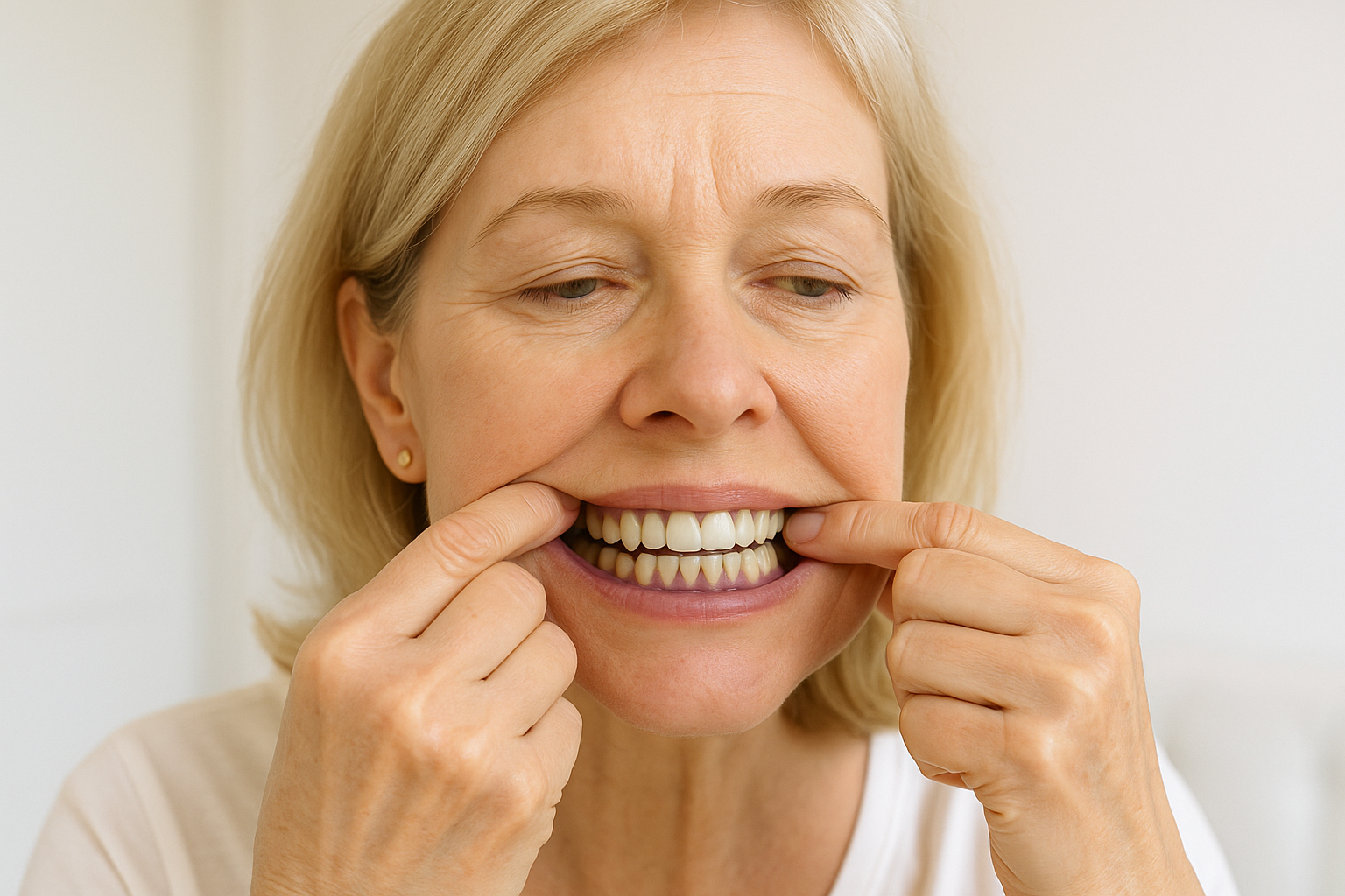
- Photograph in daylight, not the bathroom light. Use the phone’s normal camera, no filter. Pull the lip back and take a front view, left and right views, and a close-up of the darkest spot.
- Put a coin or a ruler next to the spot in the close-up, so size can be compared later.
- Write down the four sort questions: Has it always been there? Is it one spot or mouth-wide? Is it flat or swollen and shiny? Did it start after a new medicine, a dental procedure, or a change in smoking?
- For 14 days, clean the gum line properly: soft brush angled into the gum line twice a day, cleaning between the teeth daily. (Routine: how to brush with gum disease.)
- Re-photograph on day 14 in the same light with the same coin. Inflammation-colour should look calmer, less swollen and less shiny; pigment will look the same. The Cleveland Clinic’s two-week line for swollen gums is the benchmark: if they last longer than two weeks or get worse, contact a dentist.
- Book the dentist regardless if the spot is single, asymmetric, growing, ulcerated, bleeding or painful — do not wait for day 14. Show both sets of photos; they are far more useful than a description.
Why photographs: the dentists’ question is always “has it changed?”, and memory is a poor instrument for a slow change in a place you cannot see easily.
What to expect at the dentist
For pattern 1, the visit is a periodontal assessment — measuring pockets and bleeding — and a cleaning plan. For flat pigment, the dentist will usually look at the distribution, ask the four sort questions and check your medicines and history; an X-ray may be used if an amalgam tattoo is suspected. For a focal pigmented lesion that local causes do not explain, the review literature recommends biopsy, which is a small sample examined by a pathologist — it is how a benign spot is confirmed as benign. For widespread darkening plus feeling unwell, the route is via your GP for blood tests.
Where a gum-line lipid concentrate fits — and where it doesn’t
Be precise about the role, because most of the patterns above are outside what any cosmetic can address. Nothing you apply at home changes natural melanin, a smoker’s stain, an amalgam tattoo or a medicine-related pigment, and no product replaces having a single dark spot assessed. The place a daily gum-line product fits is pattern 1: while you do the cleaning that decides how the gums look, it supports the appearance of the gum tissue.
Dental Pro 7 is a 100% water-free botanical lipid concentrate built on a design principle we call Lipid-Lock: because it contains no water, it clings to the gum line and stays in contact for hours rather than being flushed away in seconds the way a water-based paste or rinse is. It blends eleven botanical lipids — immortelle helichrysum, pomegranate seed, black cumin seed, Indian myrrh, wild clove, white thyme and eucalyptus among the actives, a peppermint–spearmint–wild-mint freshness trinity, and a grapeseed, sunflower and vitamin E base — and supports the look of firmer, pinker, healthier-looking gums, with the look of redness reduced, and a mouth that looks and feels cleaner and fresher.
How to use it: four drops on a dry toothbrush in place of toothpaste, brush gently for about two minutes with the bristles angled into the gum line, then spit — do not rinse with water. Rinsing washes the lipid layer straight off and defeats the design. It suits the 14 days in the photo test above, as a companion to interdental cleaning, not a substitute for it.
Because the formula is anhydrous it is preservative-free by design — no water, no fillers, no SLS or foaming agents, no fluoride, no parabens; vegan and non-GMO. It naturally contains eugenol, limonene and linalool from its botanicals, and it is made in a facility that handles nut oils.
- Rating: Dental Pro 7 is rated 4.81 out of 5 from 190 verified buyers, with over 500,000 bottles sold.
- Guarantee: backed by a 90-day money-back guarantee.
- Formulated by S. C. Aris.
- What it is for: supporting the appearance of a cleaner, fresher mouth and healthier-looking gums. It is a cosmetic complement to brushing, interdental cleaning and professional dental care — not a substitute for any of them, and it does not lighten or change pigmentation.
Explore Dental Pro 7. Companion reading: why do my gums look like an orange peel?, what the start of receding gums looks like, and can gum disease go away?
Frequently asked questions
Why are my gums purple? Most often because they are inflamed: Cleveland Clinic says swollen gums may look reddish or purple and unusually shiny and smooth, and Mayo Clinic lists dark red or darker-than-usual gums as a gingivitis symptom. Flat purple-brown or blue-grey colour that is not swollen points to pigment instead, so the photo test above is the way to sort them.
Are dark gums a sign of gum disease? Not necessarily. Dark, swollen, shiny gums that bleed easily fit gum inflammation. Flat, even, symmetrical brown gums that have been there for years are most often natural melanin, which is more common in people with darker skin but occurs in everyone.
Can smoking make gums dark? Yes. Smoker’s melanosis is brown pigment on the gums, most often the front lip-side gums; one review reports it in 21.5% of smokers, and as few as five to nine cigarettes a day were enough in the data it summarised. It is associated with a reduction after cutting back or quitting, though time frames are not well defined.
What is a blue-grey spot on my gum near a filling? Commonly an amalgam tattoo — harmless particles of filling material in the gum — which dental reviews describe as a flat blue-grey spot, most often on the gum or the mucosa over the jaw. Because a flat dark spot can also be something else, dentists usually confirm it with an X-ray or biopsy instead of assuming.
Can a medicine turn gums dark? Yes. DermNet lists blue-black gum and tongue discolouration from some drugs, including chloroquine, hydroxychloroquine and minocycline. It mostly fades after stopping, but can be prolonged or permanent. Tell your prescriber and dentist; do not stop a prescribed medicine yourself.
When are dark gums an emergency? Rarely an emergency, but a single new dark patch that is asymmetric, growing, ulcerated, bleeding or painful should be assessed by a dentist or doctor within days, not weeks. Oral melanoma is rare, but dental literature recommends biopsy for focal pigmented lesions that local causes cannot explain.
Will a gum product make dark gums lighter? No. No cosmetic changes natural or drug-related pigment. What a gum-line product can do is support the look of healthier, pinker gums when the darkness is from inflammation, alongside proper cleaning and dental care.
The bottom line
Darker or purple gums split into colour that moves and colour that stays. Swollen, shiny, bleeding gums that are darkest along the gum line are inflammation, and they should look calmer after two weeks of thorough cleaning — and see a dentist if they do not, or sooner if painful. Flat, even, symmetrical colour is usually natural melanin, smoking or a filling-related tattoo, and needs confirming rather than treating. Colour that follows a new medicine or comes with fatigue and weight loss needs a prescriber or doctor. And a single, changing, irregular or sore dark patch is the one to get looked at this week. Take the photos; they are the best evidence you can bring.
For the wider picture, see gingivitis: how to reverse it before it becomes permanent, how gum disease is treated, and understanding puffy gums.
This article is for general information and describes cosmetic products by appearance and feel; it is not medical or dental advice. A new, single, changing, ulcerated, bleeding or painful dark patch in the mouth, or gum darkening with unexplained fatigue or weight loss, should be assessed by a dentist or doctor.