Why Is My Scar White? Pale, Depigmented or Something Else — the 3-Question Check, and What Colour Can Realistically Come Back
Quick answer
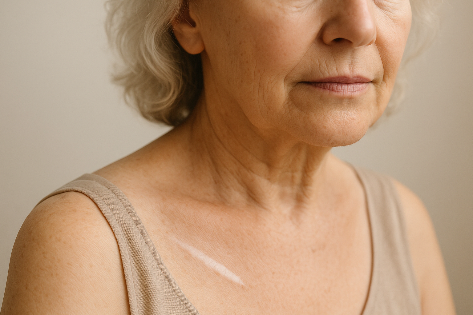
A scar looks white because the pigment cells (melanocytes) in that patch of skin were damaged or lost when the injury happened, so the repaired skin makes less melanin than the skin around it — or none. Cleveland Clinic defines hypopigmentation simply as “a low amount of melanin in your skin.” Two things decide what happens next: how much pigment is missing (pale versus chalk-white) and how long the scar has had to mature. Partial paleness in a young scar can improve as the scar matures over 12 months or more; a flat, milk-white, glossy scar a year or more on is unlikely to refill by itself, and clinical options such as lasers, grafting or medical tattooing give partial, sometimes short-lived results. Two practical rules apply either way: protect it from the sun with a broad-spectrum SPF 30+, and have a clinician look if the white area spreads beyond the scar’s own outline. A 100% water-free botanical lipid concentrate such as ScarDerma Pro is for the surface of a fully closed scar — supporting a smoother, softer-looking finish — not for putting colour back.
Last updated: 6 October 2026
Most pages on this question skip the first thing you need to know
Search “why is my scar white” and you will mostly find pages promising ways to “re-pigment” a scar with oils and home remedies. None of the clinical sources used for this article describes a topical, cream or oil that restores pigment to a scar. What the sources do describe is a short list of clinical procedures with modest, uneven results — and an equally important list of white patches that only look like scars. So this article does three things: tells you why scars go white, gives you a 3-question check to sort a true white scar from a look-alike, and puts honest numbers on how much colour can come back.
Why a scar goes white: the pigment cells took the hit
Skin colour comes from melanin, made by melanocytes in the lowest layer of the epidermis. When an injury is deep or inflamed enough to damage those cells, the skin that repairs the wound can come back with fewer of them, or with ones that no longer work properly. Thomas Mustoe, in the scar-classification chapter of the open-access Textbook on Scar Management (Springer, 2020), puts the outcome in one line: “The color of the scar ranges from white (absence of melanocytes) to hyperpigmented.” White is one end of a normal range of mature-scar colour, not a separate disease.
StatPearls’ entry on hypopigmented macules gives the mechanism in a sentence: trauma or chemical exposure causes “melanocyte injury resulting in decreased melanin.” It also draws a line that matters for your scar: hypopigmentation is decreased melanin, while depigmentation is the complete absence that follows significant loss of melanocytes. DermNet NZ uses the same split — leukoderma is “a localised area of white depigmented skin due to total loss of epidermal melanin,” and it lists scarring after severe dermatitis, infections, burns and procedures such as cryotherapy, chemical peels and lasers among the causes.
The scar-management textbook’s chapter on pigmentation disorders (Pijpe and colleagues, 2020) defines hypopigmentation the same way — skin that is lighter than the person’s own colour “but not completely lacking pigment” — and notes that it is a frequent consequence of burn injuries. That gives you a working vocabulary:
| Term | What it means | What it looks like |
|---|---|---|
| Hypopigmented | Reduced melanin — some pigment remains | Lighter than your normal skin; pink-beige, pale tan or “washed out”; the shade of your skin, diluted |
| Depigmented | No melanin — complete loss | Chalk, milk or paper white, usually smooth and sometimes slightly glossy |
Most “white scars” people describe are somewhere between the two. The distinction is not academic: it is the main thing that decides whether colour can return, as the clocks below show.
The 3-question check: true white scar, or a look-alike?
A white mark is not always a scar’s own pigment loss. DermNet’s leukoderma page groups the causes of localised white skin into scarring, autoimmune conditions (vitiligo, lichen sclerosus, systemic sclerosis, morphoea), chemical or occupational causes, congenital conditions and others, and says diagnosis relies on clinical presentation plus examination under a Wood lamp, sometimes with dermoscopy or biopsy. You cannot do those at home, but three questions will tell you whether to relax or book an appointment. This is our own triage, built from what those sources describe — an orientation aid, not a diagnosis.
- Does the white stay inside the scar’s outline? A true pigment-loss scar is white along the line or patch where the skin was actually injured, with the same edges as the injury. White that creeps beyond the edge of the scar, or new white patches appearing elsewhere, is not explained by the scar.
- Has it been stable for months? Mature scars change slowly. A white area that keeps enlarging, or new patches in other places, needs a clinician. StatPearls’ entry on the Koebner phenomenon notes that vitiligo is a condition in which new lesions can appear at sites of skin injury, and that the presence of this response correlated with higher disease activity in the preceding 12 months.
- Is the skin itself texturally different, tight, shiny or thin — beyond the scar line? DermNet lists lichen sclerosus and morphoea among the autoimmune causes of white skin; Cleveland Clinic lists lichen sclerosus among the reasons to see a provider about hypopigmentation. Skin that is white and thin, crinkled or tight over an area larger than the original wound deserves an expert look.
| What you see | Most consistent with | What to do |
|---|---|---|
| White follows the scar’s exact outline; edges unchanged for months | Pigment loss within the scar (hypopigmented or depigmented scar) | Sun protection; photo log (below); clinical options if it bothers you |
| White spreads beyond the scar edge, or new white patches appear on other areas or other scars | Not explained by the scar alone — vitiligo is one possibility that can appear at injury sites | Have a dermatologist look; do not wait for it to “settle” |
| Skin around the scar is white, thin, crinkled or tight over a wider area | A separate skin condition such as lichen sclerosus or morphoea is among the possibilities | Dermatologist assessment |
| Silvery-white, slightly sunken, wrinkled lines that followed growth or pregnancy | Striae albae — white stretch marks (see below) | Different rules; see the stretch-mark section |
What about white stretch marks?
White stretch marks are the same story on a larger canvas. StatPearls’ “Striae Distensae” entry (Mikes, Oakley and Patel, updated May 2025) describes striae albae as “hypopigmented, atrophic, and wrinkled scars” — the mature stage that appears as the earlier red stage fades over months to years — and reports that dermoscopy shows “increased melanization in striae rubrae and decreased melanization in striae albae.” So a red stretch mark and a white one are the same mark at different points in its life, and the white one has lost pigment. (Our piece on acids and stretch marks explains why the red-versus-white stage matters for what you do: can you use glycolic acid on stretch marks?)
Will the colour come back? Three clocks
The sources give different answers because they are talking about different amounts of pigment loss. Here is each clock, with the figures exactly as the sources give them:
| Clock | What the source says | What it means for your scar |
|---|---|---|
| Mild loss after a minor injury | Cleveland Clinic: for injury-related hypopigmentation, it “will usually go away after a few weeks or months” as skin heals | A pale patch after a blister or graze is often temporary — give it time before deciding it is permanent |
| Maturing scar | Mustoe: scar strength peaks at about 6 months, but “complete scar maturation as measured by resolution of erythema typically takes a year or even longer.” Pijpe et al.: hypopigmentation “often improves over time with scar maturation,” though lasting lesions “may remain” | Judge a scar’s colour at 12 months or later, not at 12 weeks — but understand that improvement is not guaranteed |
| Established pigment-loss scar | Cleveland Clinic: “If you have albinism or hypopigmentation from scars, it’s permanent,” and it states there are no effective treatments for scars that have hypopigmentation | A chalk-white scar a year or more old should not be expected to refill by itself |
| White stretch marks (striae albae) | StatPearls: broadband UV can induce repigmentation, but “this effect is typically temporary, with loss of pigmentation within a few months” | Tanning is not a fix — it fades, and it exposes skin you want to protect |
Reading the sources together — this is our synthesis, not a quoted finding — the most defensible expectation is graded. A scar that is partly pale, and still inside its first year, can improve. A scar that is completely white, flat and a year or more old has most likely settled where it is. Cleveland Clinic is blunter than the textbook, and the difference comes down to how much pigment is left to work with.
What clinical options exist, and how well do they work?
Honest answer: the options are real but limited. Two recent reviews state this plainly.
Lasers. Baugh, Anagu and Kelly’s 2022 review in Dermatologic Surgery opens with the problem: “repigmentation of hypopigmented scars remains a difficult clinical problem.” Their conclusions: “Reliable improvement of hypopigmentation in scars after laser treatment is challenging.” Ultraviolet laser “can achieve modest repigmentation; however, results are short-lived and require continued re-treatment,” and the best results came from combining fractional laser resurfacing with a topical prostaglandin analogue, with or without a topical retinoid. Those are prescription, clinician-directed treatments.
Surgical and cell-based approaches. The pigmentation chapter in the scar-management textbook (Pijpe et al., 2020) lists excimer laser (308 nm) therapy, medical tattooing and camouflage products as non-surgical options, and dermabrasion with melanocyte transplantation, skin grafting, autologous epidermal cell suspensions, microneedling as an adjunct, and excision as surgical ones — and advises trying the conservative options first. A 2018 review by Carney and colleagues in Plastic and Reconstructive Surgery — Global Open notes that, unlike excess pigmentation, skin-darkening strategies for hypopigmentation in burn scars have received far less attention; it examines melanocyte-keratinocyte transplantation, synthetic alpha-melanocyte-stimulating hormone analogues and FK506 as routes, and says laser-assisted drug delivery is a direction for future research.
The practical takeaway: if the colour difference bothers you, the right route is a dermatologist or plastic surgeon who treats scar pigmentation — and camouflage (colour-matched cosmetic cover, or medical tattooing in the hands of an experienced practitioner) is a legitimate, immediate option the sources themselves list. No cream, oil or serum, including ours, appears in the clinical literature as a way to restore a scar’s lost pigment.
Sun protection for a white scar
Cleveland Clinic’s advice on hypopigmented skin is to use sunscreen with an SPF of at least 30 to protect the affected areas. Our own reasoning, not a cited finding: skin with little or no melanin has less of its natural UV buffer than the skin around it, and a scar is a patch you want to look after anyway. In practice that means a broad-spectrum SPF 30+ on exposed scars once they are fully closed, reapplied, plus clothing or shade for areas such as the chest, shoulders and legs. This is the opposite of the brown-scar problem — there, sun darkens the scar (see why is my scar dark?) — but the habit is the same.
The 12-month colour log
The sources do not offer a way to measure progress at home, which is the gap this fills. It is our protocol, built to answer the one question that matters: is the white area stable, shrinking, or spreading?
| When | Do | What it tells you |
|---|---|---|
| Day 1 | Photograph the scar next to a plain ruler, in daylight by a window, no flash, same distance each time. Note the date | Your baseline |
| Every month | Repeat from the same angle and distance, at the same time of day. Do not edit or filter the photos | Whether the edge of the white area is moving |
| Month 3 and month 6 | Put the photos side by side. Is the colour closer to your skin tone, the same, or paler? | Whether the scar is still maturing in your favour |
| Month 12 | Compare with day 1. If it is unchanged and bothers you, book an appointment with the pictures in hand | Whether to treat it as settled — and a record to show a clinician |
| Any time | If white appears beyond the scar edge, or on other scars or areas of skin, book sooner | The signal from question 1 and 2 above |
When to see a clinician
- The white area is spreading beyond the original scar, or new white patches are appearing elsewhere.
- The white skin is also thin, tight, itchy or crinkled over an area larger than the original injury.
- The scar is painful, newly raised, ulcerated or changing shape — colour is not the concern then.
- The colour difference is affecting how you feel about the area. Cleveland Clinic lists exactly that — the condition affecting your mental health — as a reason to talk to a provider, and the options above exist for it.
Where a water-free lipid concentrate fits: ScarDerma Pro
Given what the sources say, the honest job for a topical on a white scar is narrow: look after the surface of skin that has fully closed, and support the look of a smoother, softer finish. It does not replace sun protection, and it is not a way to restore lost pigment — nothing on a shelf is, according to the clinical literature above.
ScarDerma Pro is a 100% water-free (anhydrous) botanical lipid concentrate, formulated by S. C. Aris. Because it contains no water, it needs no preservative system, and its lipids are chosen to sit in the outer layers of the skin rather than evaporating away. The formula leads with four actives — immortelle helichrysum, rosehip seed, tamanu and pomegranate seed — with Japanese camellia, geranium, English lavender and hemp seed as soothers, on a lipid base of sea buckthorn, calendula, jojoba, hazelnut and vitamin E. There are no harsh acids or peels, no drying agents, no artificial fragrance, no parabens and no fillers; it is vegan and non-GMO. One honest caveat: helichrysum, geranium and lavender are aromatic botanicals, so this is not a fragrance-free product — patch test on unbroken skin near the scar first (method in why does my serum sting?).
Its purpose is cosmetic — supporting the appearance of smoother, softer, more even-looking scars and stretch marks once the skin is fully closed. In a customer survey, 91% said their marks looked flatter and smoother within six weeks, and 94% would recommend it; these are survey responses, not clinical-trial results, and the survey did not measure pigment return. It is rated 4.91 out of 5 from 34 verified buyers and carries a 90-day guarantee. See ScarDerma Pro →
The boundary, stated plainly: use it on fully closed skin, and keep SPF 30+ running alongside it. If the white area is spreading, or the skin around it is thin or tight, the next step is a dermatologist, not a bottle.
More from our scar cluster: why is my scar dark? · why is my scar still red? · why is my scar indented? · why is my scar shiny? · why is my scar getting wider? · when can you start scar cream? · do old scars ever fade?
Frequently asked questions
Why is my scar white?
Because the pigment cells in that patch of skin were damaged or lost when the injury happened, so the repaired skin makes less melanin than the skin around it. Mustoe describes mature scar colour as ranging from white, reflecting an absence of melanocytes, to hyperpigmented. StatPearls separates reduced melanin (hypopigmentation) from complete absence (depigmentation).
Will my white scar get its colour back?
It depends on how much pigment is missing and how long it has had to mature. Cleveland Clinic says injury-related hypopigmentation usually goes away after a few weeks or months, but that hypopigmentation from scars is permanent. The scar-management textbook says it often improves with scar maturation, though lasting lesions may remain. Scar maturation itself typically takes a year or longer according to Mustoe.
Can I treat a white scar at home?
None of the clinical sources used for this article describes a cream, oil or home remedy that restores pigment to a scar. Cosmetic camouflage is a listed option for appearance, and the clinical routes (lasers, grafting, cell-based procedures, medical tattooing) are decisions for a dermatologist or plastic surgeon.
Does laser treatment fix white scars?
Partly, and not always permanently. A 2022 review in Dermatologic Surgery concluded that reliable improvement of hypopigmentation in scars after laser treatment is challenging, that ultraviolet laser can achieve modest repigmentation with short-lived results requiring re-treatment, and that combining fractional laser with a topical prostaglandin analogue gave superior repigmentation.
Do white stretch marks come back to normal colour?
White stretch marks (striae albae) are described by StatPearls as hypopigmented, atrophic, wrinkled scars. Broadband UV can induce repigmentation, but StatPearls says the effect is typically temporary, with loss of pigmentation within a few months, and notes that topical treatments are frequently recommended despite minimal evidence supporting their effectiveness.
Should I put sunscreen on a white scar?
Yes, once it is fully closed. Cleveland Clinic advises sunscreen with an SPF of at least 30 for areas of hypopigmented skin. Choose a broad-spectrum product and reapply, and cover exposed areas with clothing or shade.
How do I tell a white scar from vitiligo?
Look at where the white sits. White that stays inside the outline of the injury and has been stable for months is consistent with pigment loss in the scar. White that spreads beyond the scar, or new white patches elsewhere, is not explained by the scar, and vitiligo is one condition that can appear at sites of skin injury. Only a clinician can tell them apart, usually with a Wood lamp examination, so have it checked.
Is a white scar a sign of something wrong?
Usually not; white is part of the normal colour range of mature scars. It deserves attention if the white area is spreading, if the skin around it is thin, tight or crinkled, or if the scar is painful, raised or changing shape.
This article is general information, not medical advice, and does not replace your surgeon’s, dermatologist’s or nurse’s instructions for your own scar. The three-question check is an orientation aid, not a diagnostic test. ScarDerma Pro is a cosmetic product intended to support the appearance of healed skin; it is not intended to diagnose, treat, cure or prevent any condition, and it should only be applied to fully closed skin. See a doctor if a white area is spreading, if the skin around a scar is thin or tight, or if a scar is painful, raised or changing.
Sources
- Mustoe TA. International Scar Classification in 2019. In: Téot L, Mustoe TA, Middelkoop E, et al., eds. Textbook on Scar Management. Springer; 2020, ch. 9. NCBI Bookshelf NBK586057
- Cleveland Clinic. Hypopigmentation. Last updated 27 June 2022. my.clevelandclinic.org
- Pijpe A, Gardien KLM, van Meijeren-Hoogendoorn RE, Middelkoop E, van Zuijlen PPM. Scar Symptoms: Pigmentation Disorders. In: Textbook on Scar Management. Springer; 2020. link.springer.com
- Baugh EG, Anagu O, Kelly KM. Laser Treatment of Hypopigmentation in Scars: A Review. Dermatol Surg. 2022. DOI 10.1097/dss.0000000000003330. escholarship.org
- Carney BC, McKesey JP, Rosenthal DS, Shupp JW. Treatment Strategies for Hypopigmentation in the Context of Burn Hypertrophic Scars. Plast Reconstr Surg Glob Open. January 2018. doaj.org
- Tan K, Keefe M, Ngan V. Leukoderma. DermNet NZ. Updated August 2021. dermnetnz.org
- Hypopigmented Macules. StatPearls. statpearls.com
- Sanchez DP, Sonthalia S. Koebner Phenomenon. StatPearls. Updated 14 November 2022. NCBI Bookshelf NBK553108
- Mikes BA, Oakley AM, Patel BC. Striae Distensae. StatPearls. Updated 19 May 2025. NCBI Bookshelf NBK436005